animal cell under microscope diagram
After the invention of the microscope animal cells were first observed in the 17th century. A Dutch scientist named Anton van Leeuwenhoek observe these cells under the microscope and described bacterial cells and red blood cells of animals.

Image Result For Diagram Of Plant And Animal Cell Under Electron Microscope Celula Animal Ciencias Verdades Absolutas
Animal Cell Structure and Cell Organelles.

. Animal Cell Diagram Under Microscope. Thousands of new high-quality pictures added every day. The cell was first discovered by an English natural philosopher Robert Hooke.
Animal cell structure varies in different shapes. Under the microscope animal cells appear different based on the type of the cell. Almost all animals and plants are made up of cells.
Many of us have failed to recognise the. This is due to the lack of a cell wall. The granulated area is the cell Cytoplasm while the huge round part is the Nucleus.
Animal Cell Under Microscope. They are all typical elements of a cell. Friday May 6 2022.
The first animal cell was observed under an optical microscope which clearly showed the nucleus and microfilament network in red and blue colors respectively. Animal Cell Diagram Under Light Microscope. We use microscope comprehensively in microbiology mineralogy cell biology biotechnology nano physics microelectronics pharmacology and forensics.
Typical Animal Cell With Labels. Find Animal cell under microscope stock images in HD and millions of other royalty-free stock photos illustrations and vectors in the Shutterstock collection. While observing with tissues or on tissue.
Animal cell under microscope diagram Saturday January 29 2022 Edit. Most cells both animal and plant range in size between 1 and 100 micrometers and are thus visible only with the aid of a microscope. Observing a wide range of biological processes and animal cell under light microscope is easier due to advances in microscopic techniques.
Function cell does in the body dictate the change and adaptation done by cell. When observing onion cells there is the Cell Surface Membrane which is present in all living cells. A typical animal cell is 1020 μm in diameter which is about one-fifth the size of the smallest particle visible to the naked eye.
Examining animal cells under the microscope. We all keep in mind that the human body is quite intricate and a method I discovered to are aware of it is via the manner of. Under the microscope animal cells appear different based on the type of the cell.
With a light microscope you can see several structures inside the cell. This shows a generalized animal cell under a light. A cell is the smallest functional and structural entity of life that it is easier observing animal cell under light microscope lensclutcolunch.
There are various tasks done by a cell to complete them as the. Typical Animal Cell Pinocytotic vesicle Lysosome Golgi vesicles Golgi vesicles rough ER endoplasmic reticulum Smooth ER no ribosomes Cell plasma membrane Mitochondrion Golgi apparatus Nucleolus Nucleus Centrioles 2 Each composed of 9 microtubule triplets Microtubules Cytoplasm Ribosome. Look at the diagram which identifies the different components in a simple animal cell.
Plant and Animal. However the internal structure and organelles are more or less similar. A typical animal cell is 1020 μm in diameter which is about one-fifth the size of the smallest particle visible to the naked eye.
Animal cell under electron microscope. A cell structure that controls which substances can enter or leave the cell. As you can see in the above labeled plant cell diagram under light microscope there are 13 parts namely Cell membrane.
Human animal cell under microscope. Microscopic Organisms In A Drop Of Pond Water Microscopic Organisms Things Under A Microscope Microscopic Structure Of Animal Cell And Plant Cell Under Microscope Diagrams Cell Diagram Plant Cell Diagram Animal Cell. Animal cells have a basic structure.
Animal cell under microscope diagram Saturday January 29 2022 Edit. Animal and plant cell under electron microscope. Light and electron microscopes allow us to see inside cells.
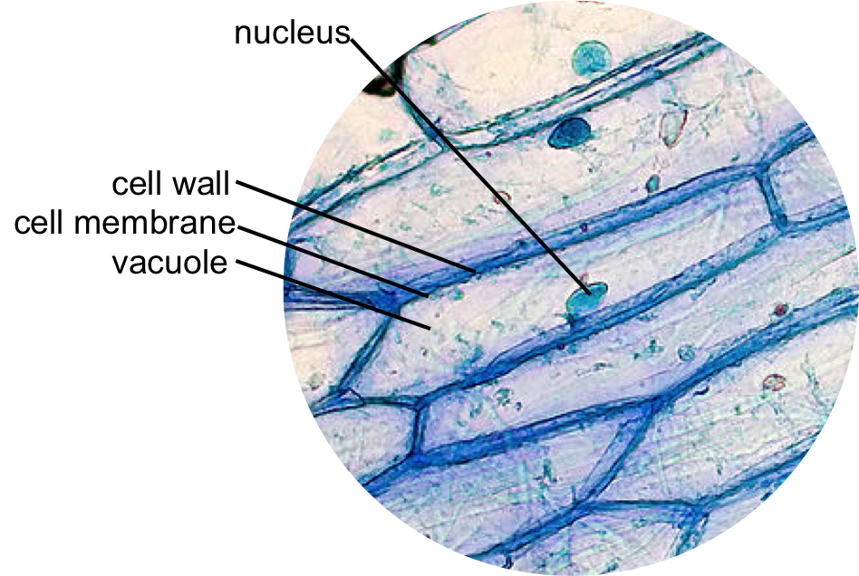
Structure of animal cell and plant cell under microscope diagrams. When observing onion cells there is the Cell Surface Membrane which is present in all living cells. The ability to distinguish clearly the individual parts of an object under a microscope.
Cell structure and organisation_notes igcsebiology dnl. One animal and one plant example given. Diagram 32 an.
Plant cells have cell walls one large vacuole per cell and chloroplasts while animal cells will have a cell membrane only. Below the basic structure is shown in the same animal cell on the left viewed with the light microscope. Onion Epidermis With Large Cells Under Light Microscope.
Animal cell under the microscope. Some may be oval or cylindrical shaped. Generalized Structure of a Plant Cell Diagram.

Animal Cell Plant Cell Diagram Cell Diagram Animal Cell

Animal Cell Organelles Cell Organelles Organelles

Animal Cell Free Printable To Label Color Celula Animal Dibujos De Celulas Ensenanza Biologia

How To Draw Animal Cell Biology Drawing Animal Cell Animal Cell Drawing

Draw It Neat How To Draw Animal Cell Animal Cell Animal Cell Drawing Cell Diagram

Animal Cell Structure And Organelles With Their Functions Animal Cell Organelles Plant And Animal Cells

Animal Cell Model Diagram Project Parts Structure Labeled Coloring Animal Cell Plant And Animal Cells Animal Cells Model

Plant Cell Diagram Animal Cell Diagram Plant And Animal Cells Animal Cell Science Cells

Cells Under Electron Microscope Google Search Animal Cell Structure Animal Cells Worksheet Cell Diagram

This Schematic Diagram Shows A Generic Animal Cell And The Organelles Including The Nucleus Endopla Human Cell Diagram Human Cell Structure Animal Cell Parts

Animal And Plant Cells Worksheet Inspirational 1000 Images About Plant Animal Cells On Pinterest Cells Worksheet Plant Cells Worksheet Animal Cell

Epidermal Onion Cells Under A Microscope Plant Cells Appear Polygonal From The Cell Diagram Plant Cell Diagram Plant Cell

Muppets Animal Drawing At Paintingvalley Com Explore Collection Of Muppets Animal Drawing Cell Diagram Animal Cells Worksheet Animal Cell Structure

Animal Cell Structure And Organelles With Their Functions Animal Cell Organelles Cell Diagram

Learn About The Plant Cell Science For Kids And Science Activities And Projects For Kids Plant Cell Cell Diagram Animal Cell Structure

Google Image Result For Http W3 Hwdsb On Ca Hillpark Departments Science Watts Sbi3u Assigned Work Cell Struct Plant Cell Cell Diagram Animal Cells Worksheet

Year 11 Bio Key Points Cell Organelles And Their Function Cell Organelles Animal Cell Organelles

Diagram Showing Anatomy Of Animal Cell Royalty Free Vector ช วว ทยาศาสตร การศ กษา คำคมการเร ยน
